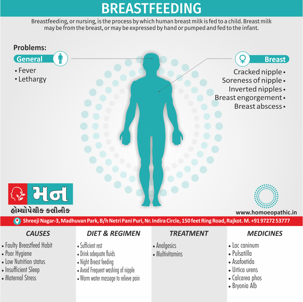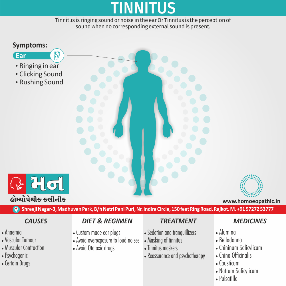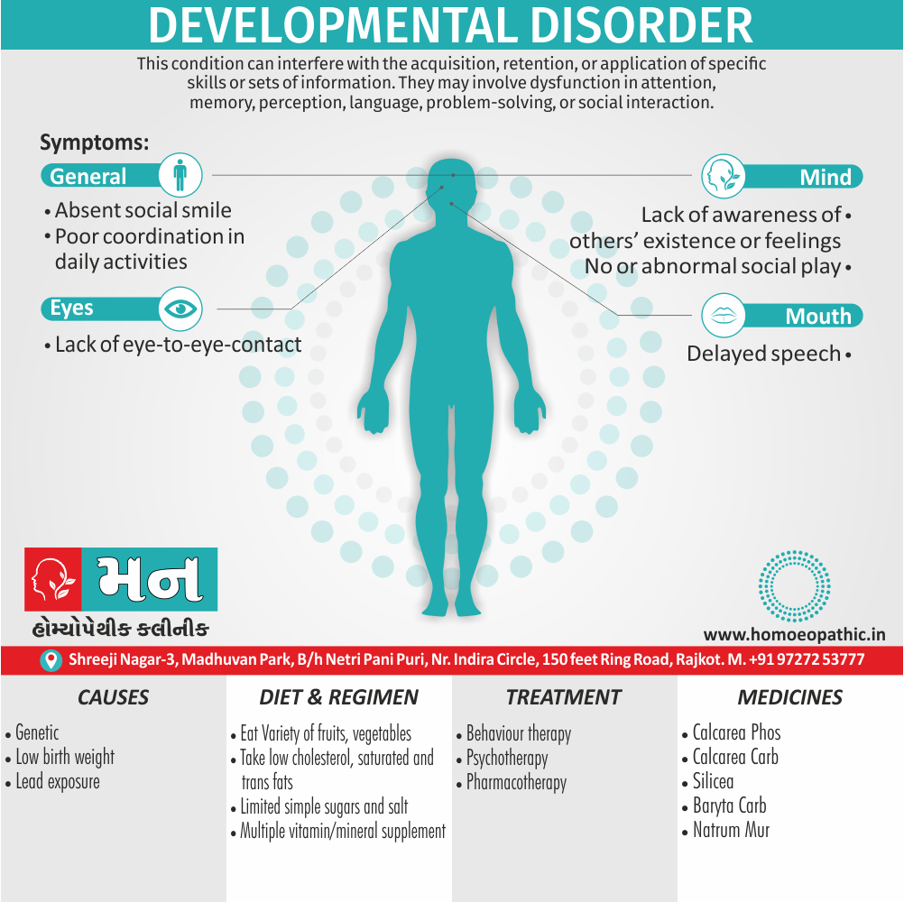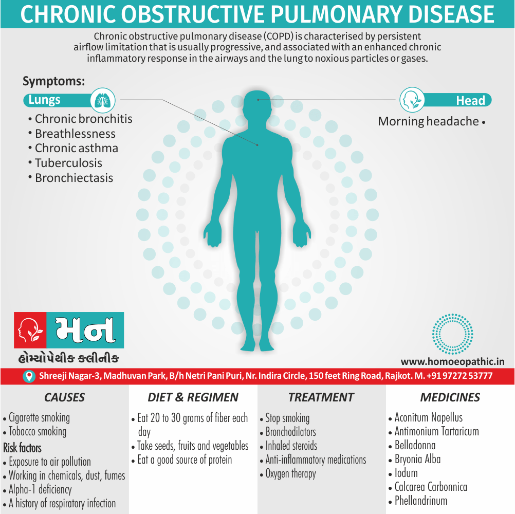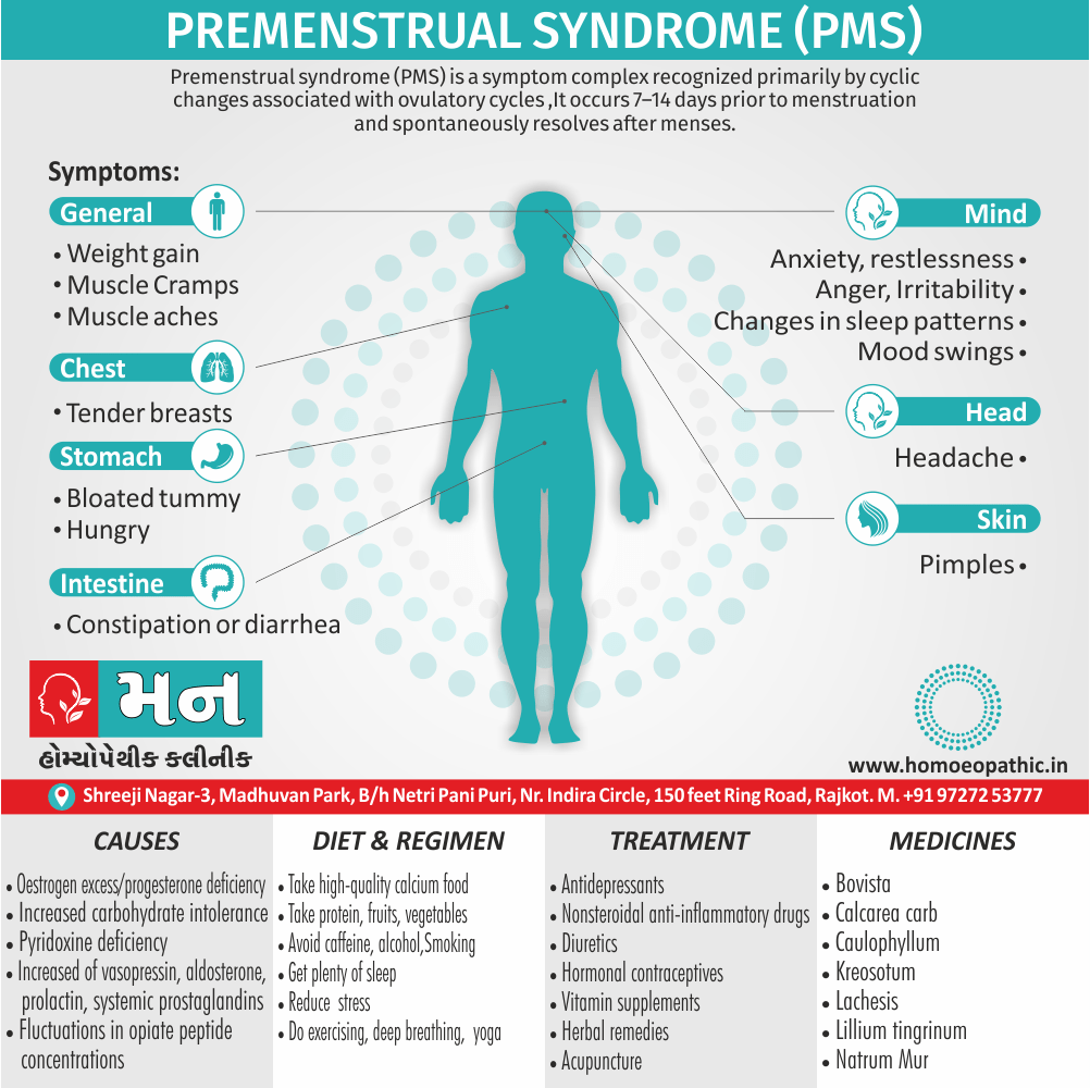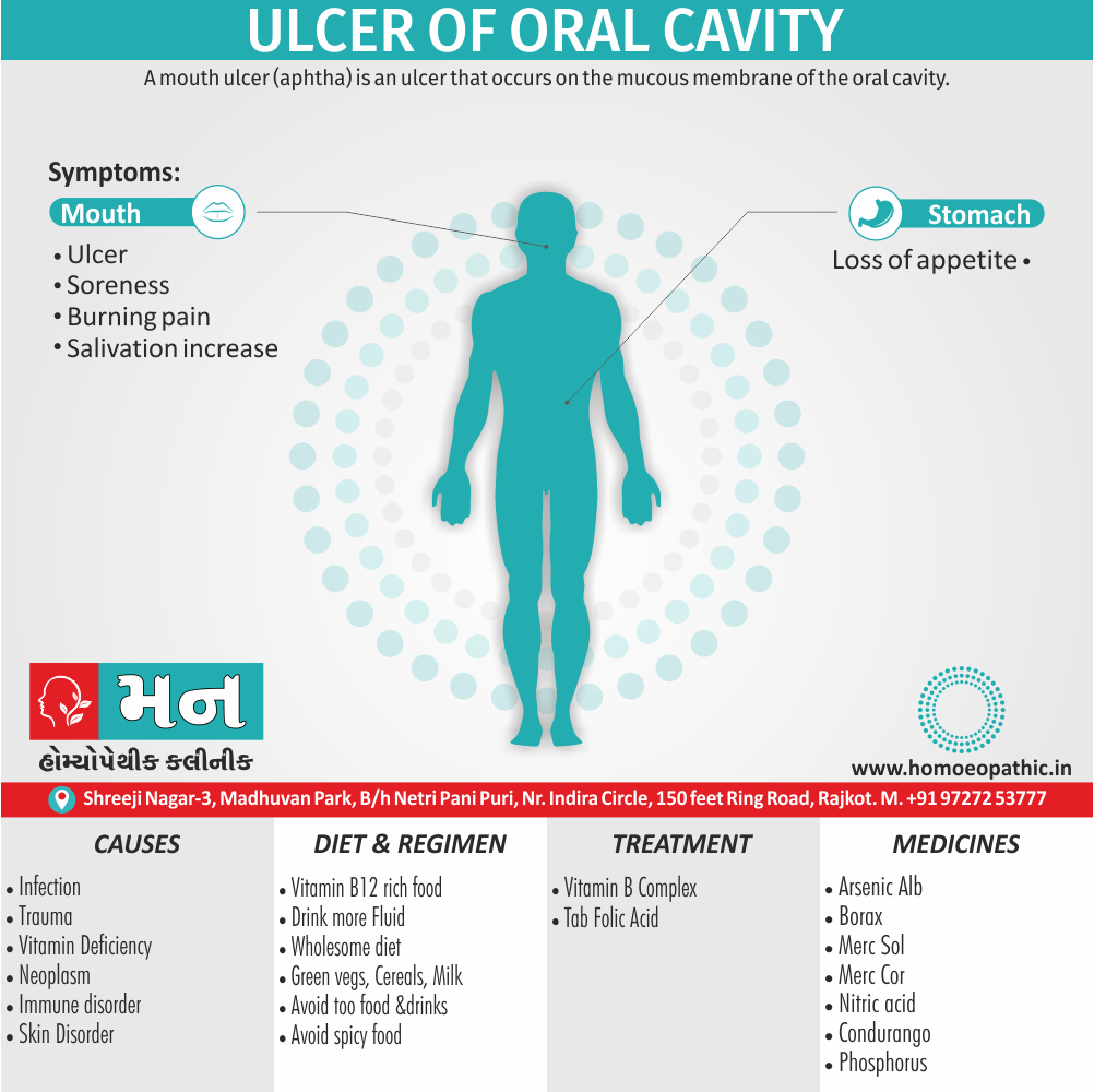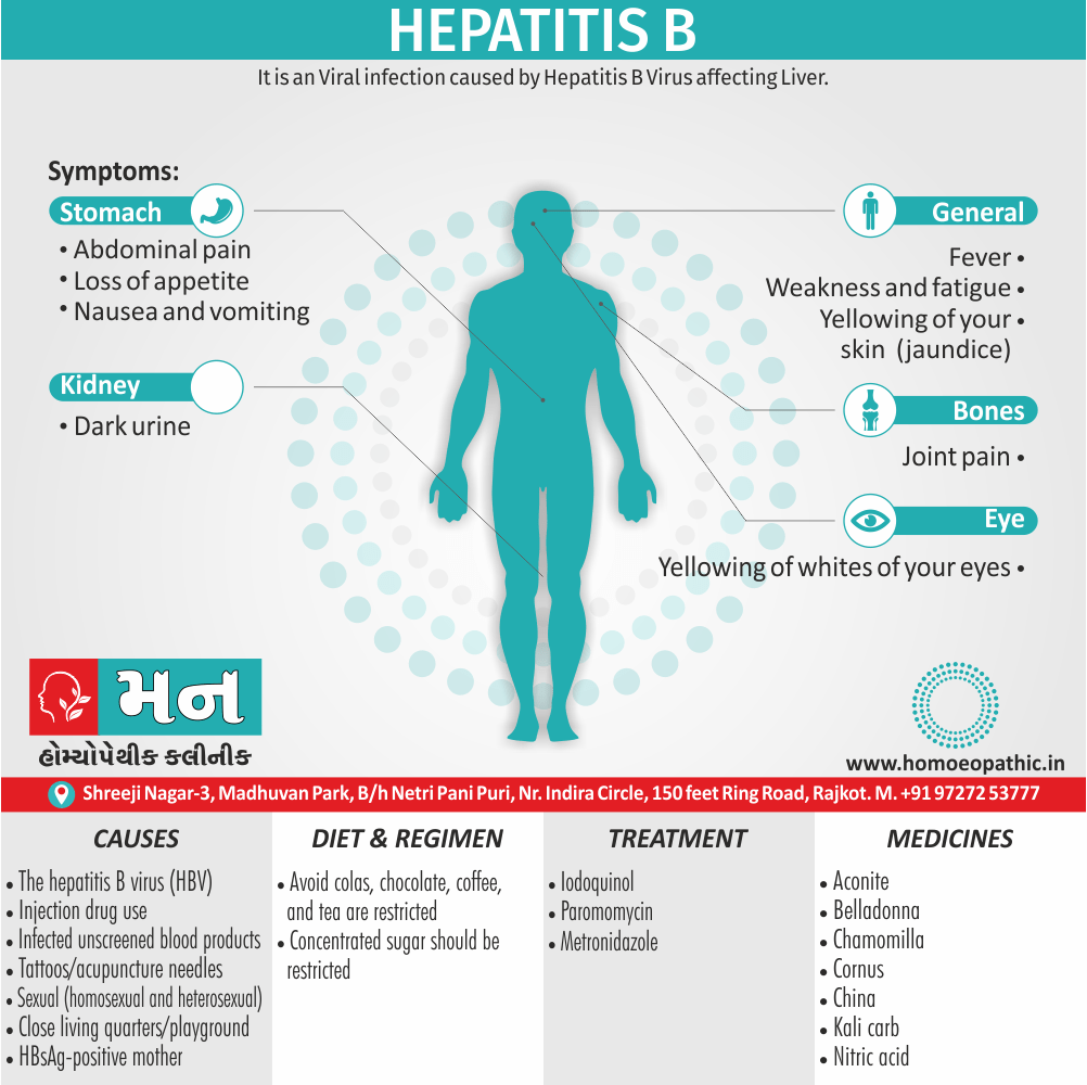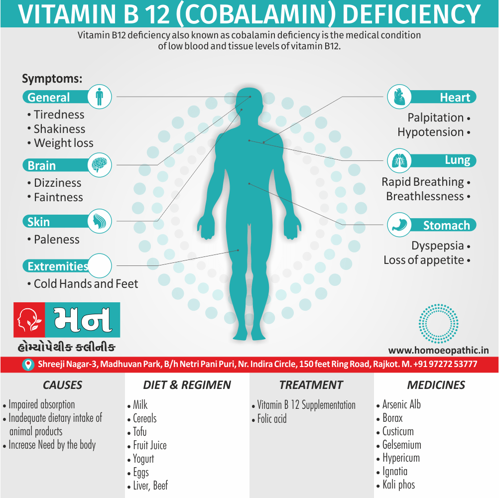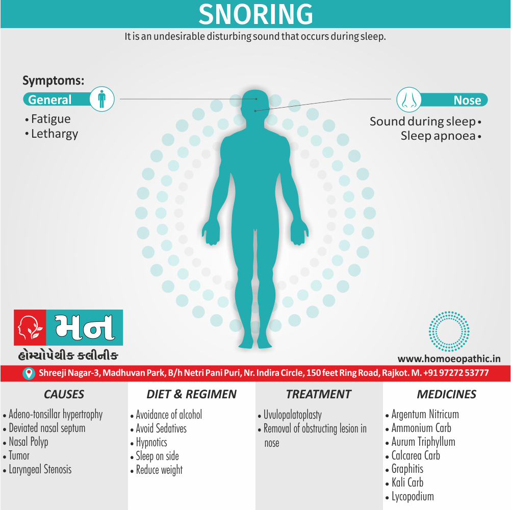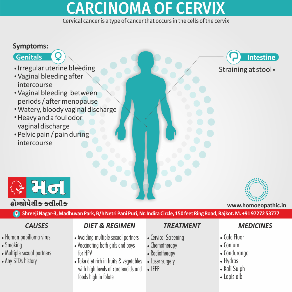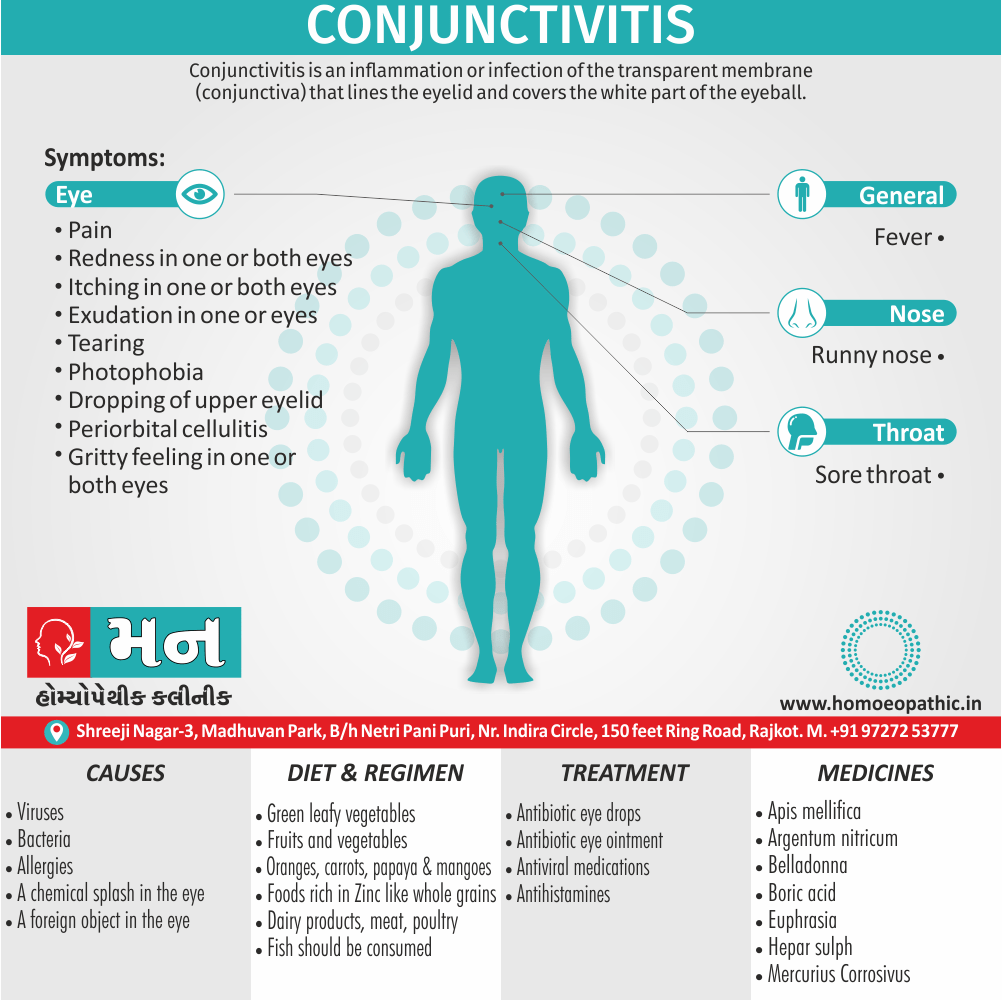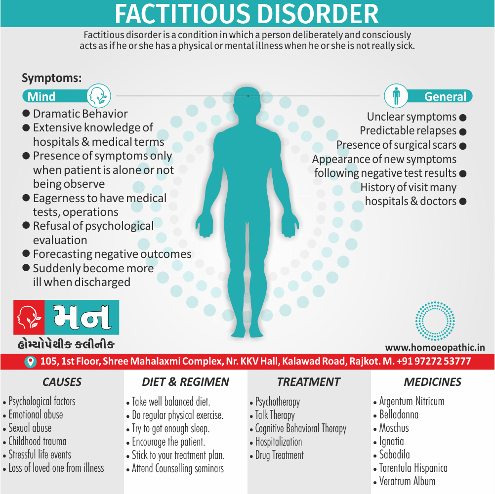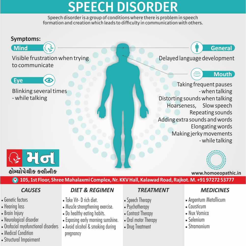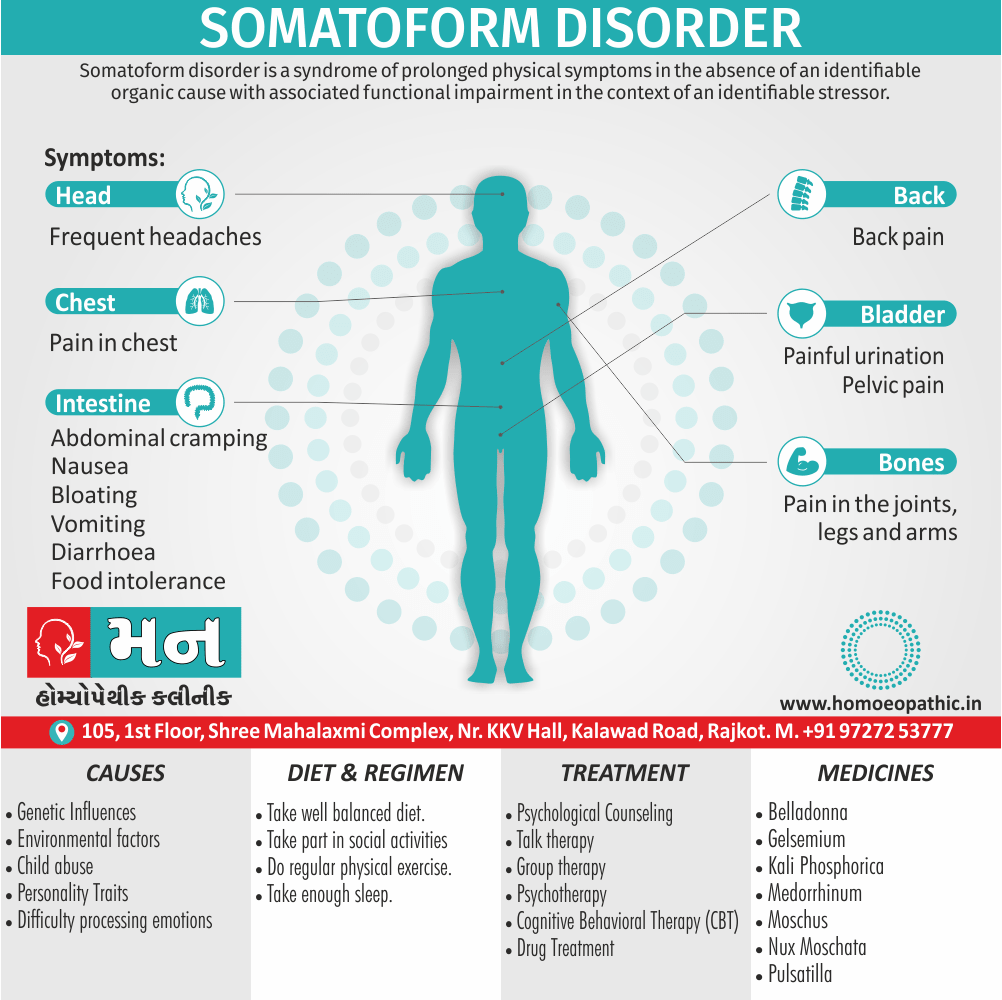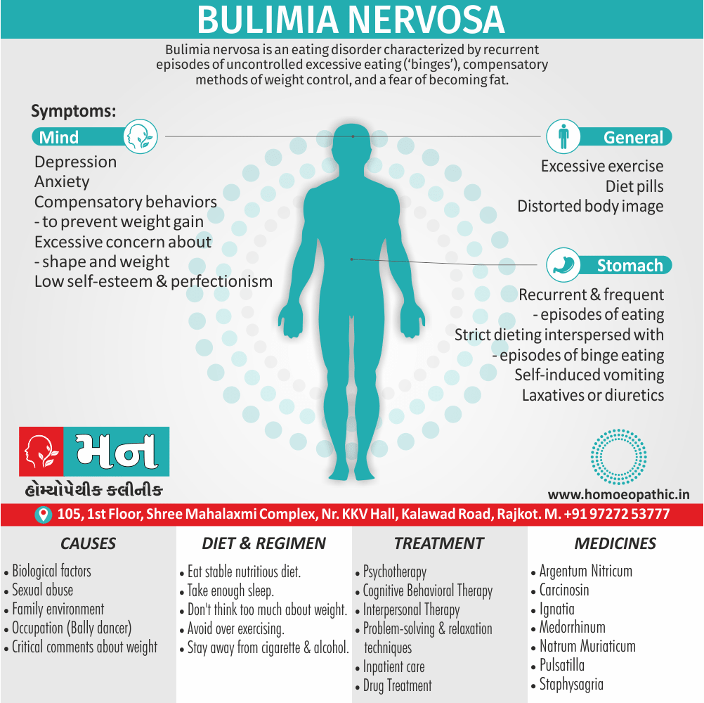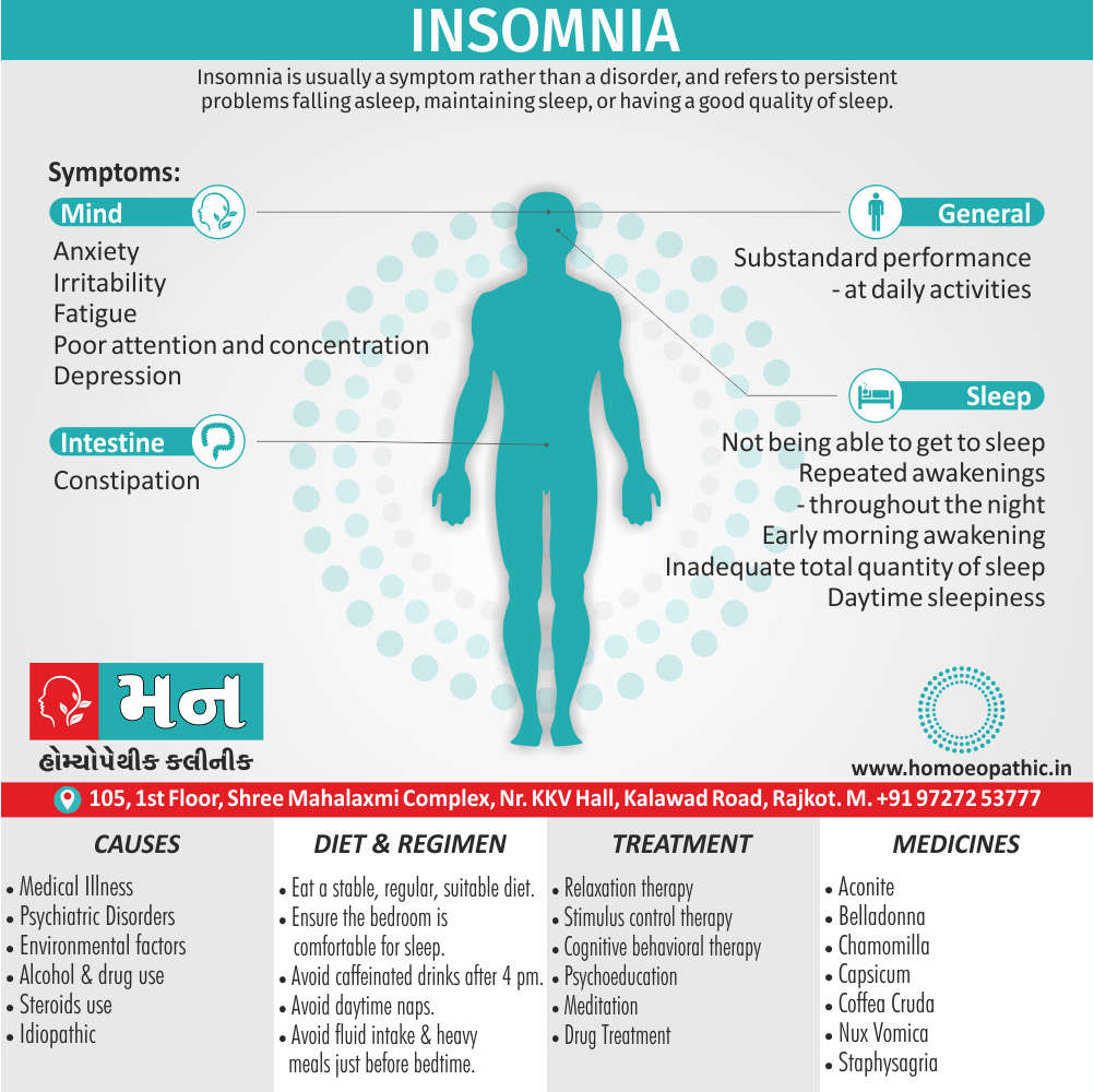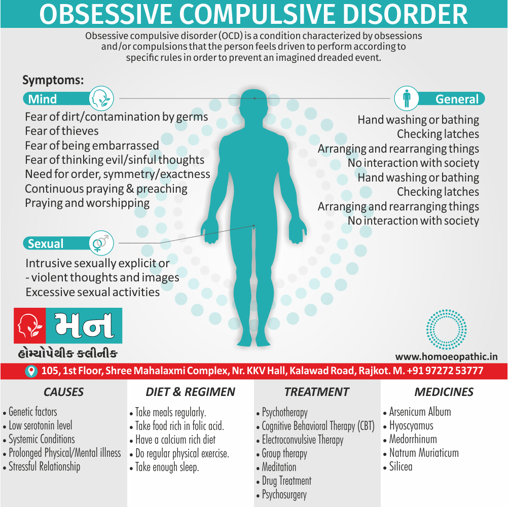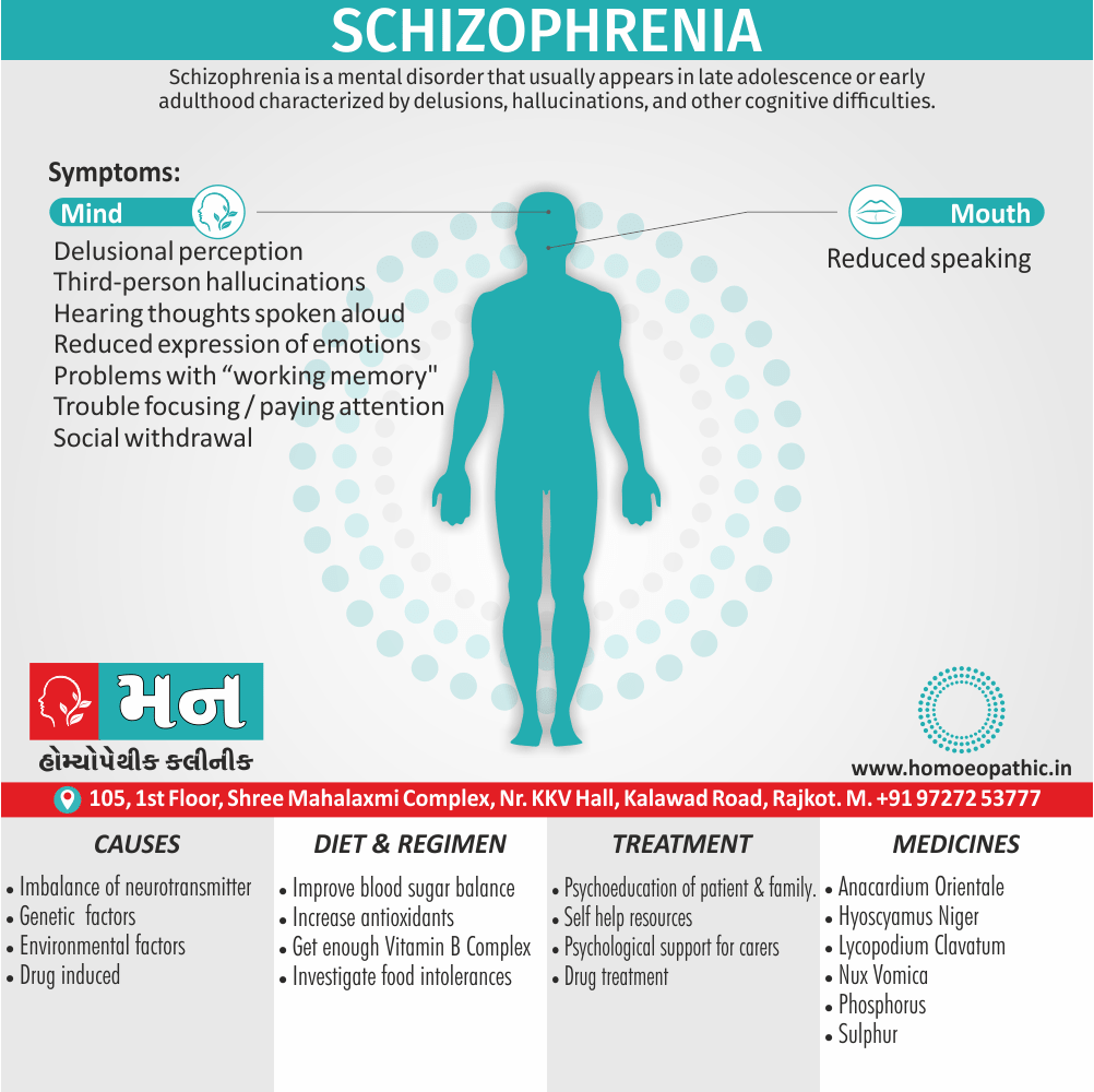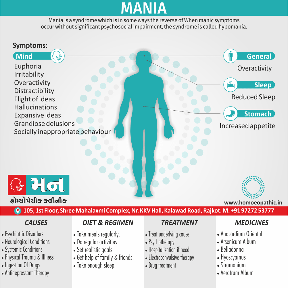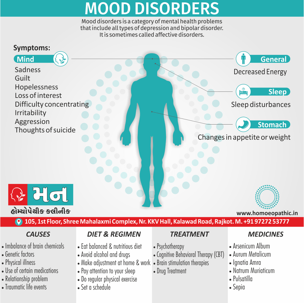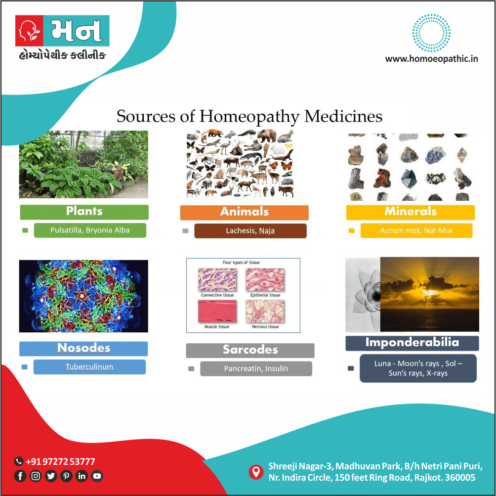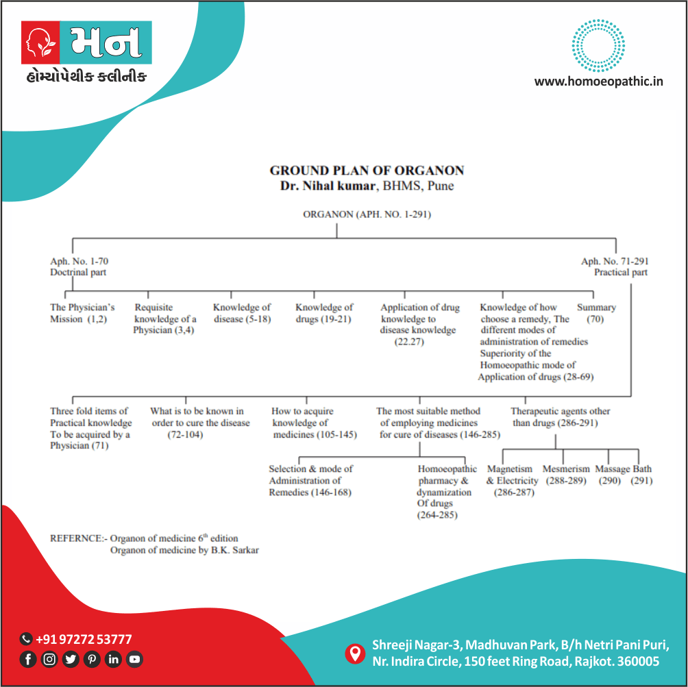
Skeletal System:
Skeletal System Consists of the axial skeleton (bones of the head, vertebral column, ribs, also sternum) and the
appendicular skeleton (bones of the extremities).
Bones and Joints are important parts of Skeletal System.
I. Bones in Skeletal System:
- Bons are calcified connective tissue consisting of cells (in other words; osteocytes) embedded in a matrix of ground substance and collagen fibers, have a superficial thin layer of compact bone around a central mass of spongy bone, also contain internal soft tissue, the marrow, where blood cells are formed.
- It serve as a reservoir for calcium also phosphorus and act as biomechanical levers on which muscles act to produce the movements permitted by joints.
- Lastly, Bons are classified, according to shape, into long, short, flat, irregular, and sesamoid bones and, according to their developmental history, into endochondral and membranous bones.
A. Long Bones
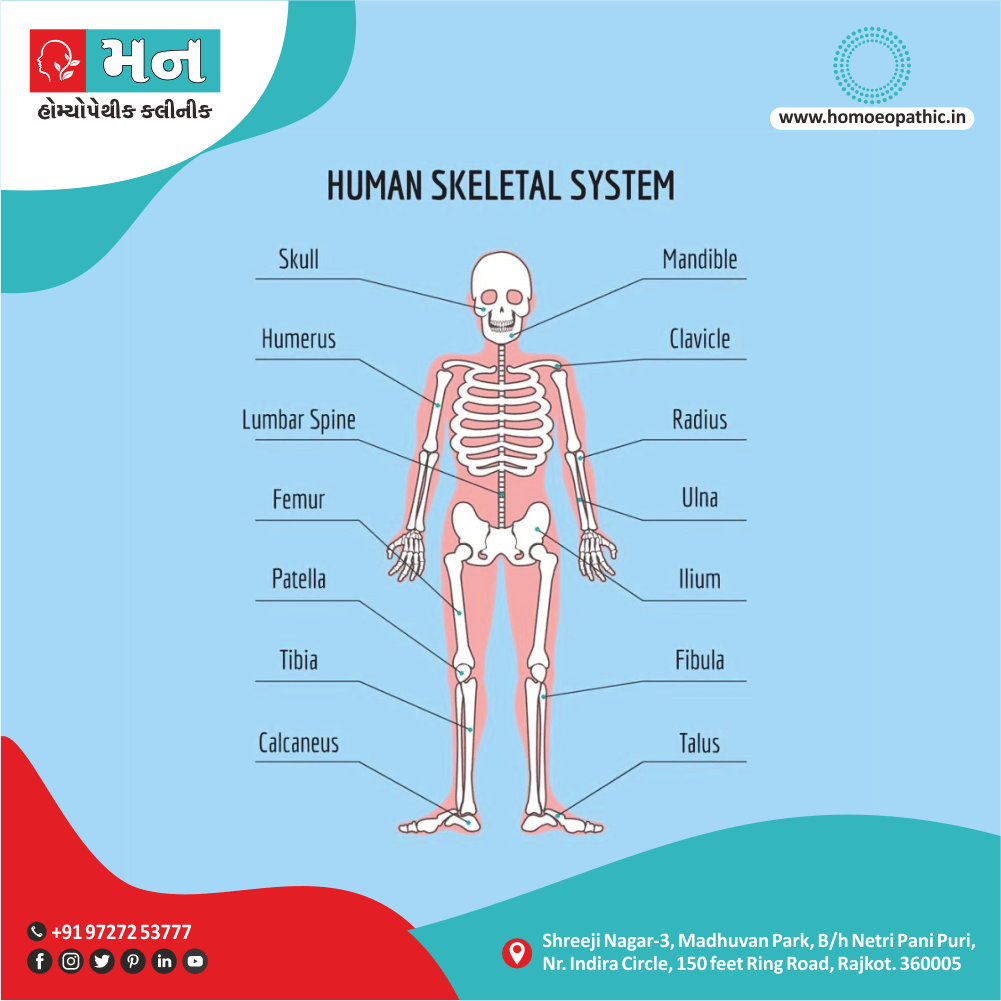
- Include the humerus, radius, ulna, femur, tibia, fibula, metacarpals, and phalanges.
- Develop by replacement of hyaline cartilage plate (endochondral ossification).
- Have a shaft (diaphysis) and two ends (epiphyses). Additionally, The metaphysis is a part of the diaphysis adjacent to the epiphyses.
1. Diaphysis i.e.
■ Forms the shaft (central region) and is composed of a thick tube of compact bone that encloses the marrow cavity.
2. Metaphysis i.e.
■ Is a part of the diaphysis, the growth zone between the diaphysis and epiphysis during bone development.
3. Epiphyses i.e.
■ Are expanded articular ends, separated from the shaft by the epiphyseal plate during bone growth and composed of a spongy bone surrounded by a thin layer of compact bone.
B. Short Bones
■ Include the carpal and tarsal bones and are approximately cuboid-shaped.
■ Are composed of spongy bone and marrow surrounded by a thin outer layer of compact bone.
C. Flat Bones
- Include the ribs, sternum, scapulae, and bones in the vault of the skull.
- Consist of two layers of compact bone enclosing spongy bone and marrow space (diploë)
- Have articular surfaces that are covered with fibrocartilage and grow by the replacement of
connective tissue.
D. Irregular Bones
■ Include bones of mixed shapes such as bones of the skull, vertebrae, and coxa.
■ Contain mostly spongy bone enveloped by a thin outer layer of compact bone.
E. Sesamoid Bones
■ Develop in certain tendons and reduce friction on the tendon, thus protecting it from excessive wear.
■ Are commonly found where tendons cross the ends of long bones in the limbs, as in the wrist and the knee (i.e., patella).
CLINICAL ANATOMY
- Osteoblast synthesizes new bone and osteoclast functions in the resorption (break down bone matrix also release calcium and minerals) and remodeling of bone. Parathyroid hormone causes mobilization of calcium by promoting bone resorption, whereas calcitonin suppresses mobilization of calcium from bone. Additionally, Osteoid is the organic matrix of bone prior to calcification.
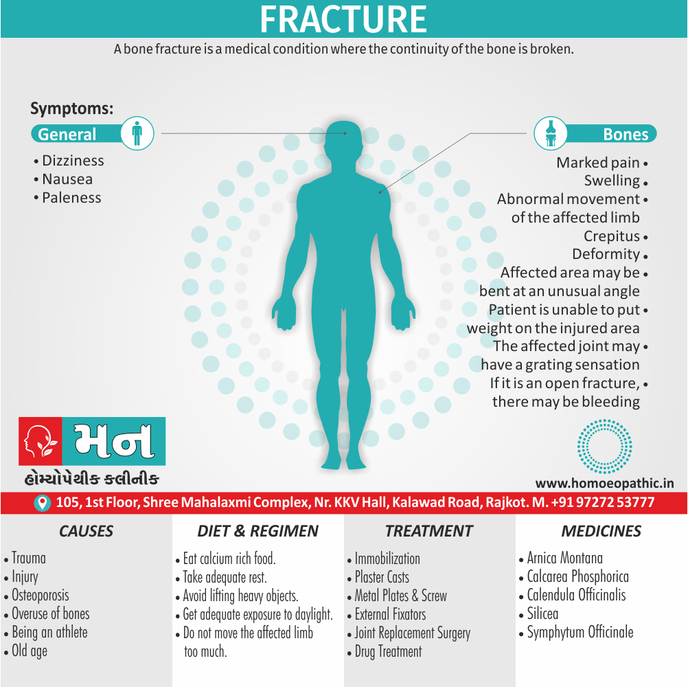
- Osteomalacia is a gradual softening of the bone due to failure of the bone to calcify because of either lack of vitamin D or renal tubular dysfunction.
- Osteopenia is a decreased either calcification of bone or a reduced bone mass due to an inadequate osteoid synthesis.
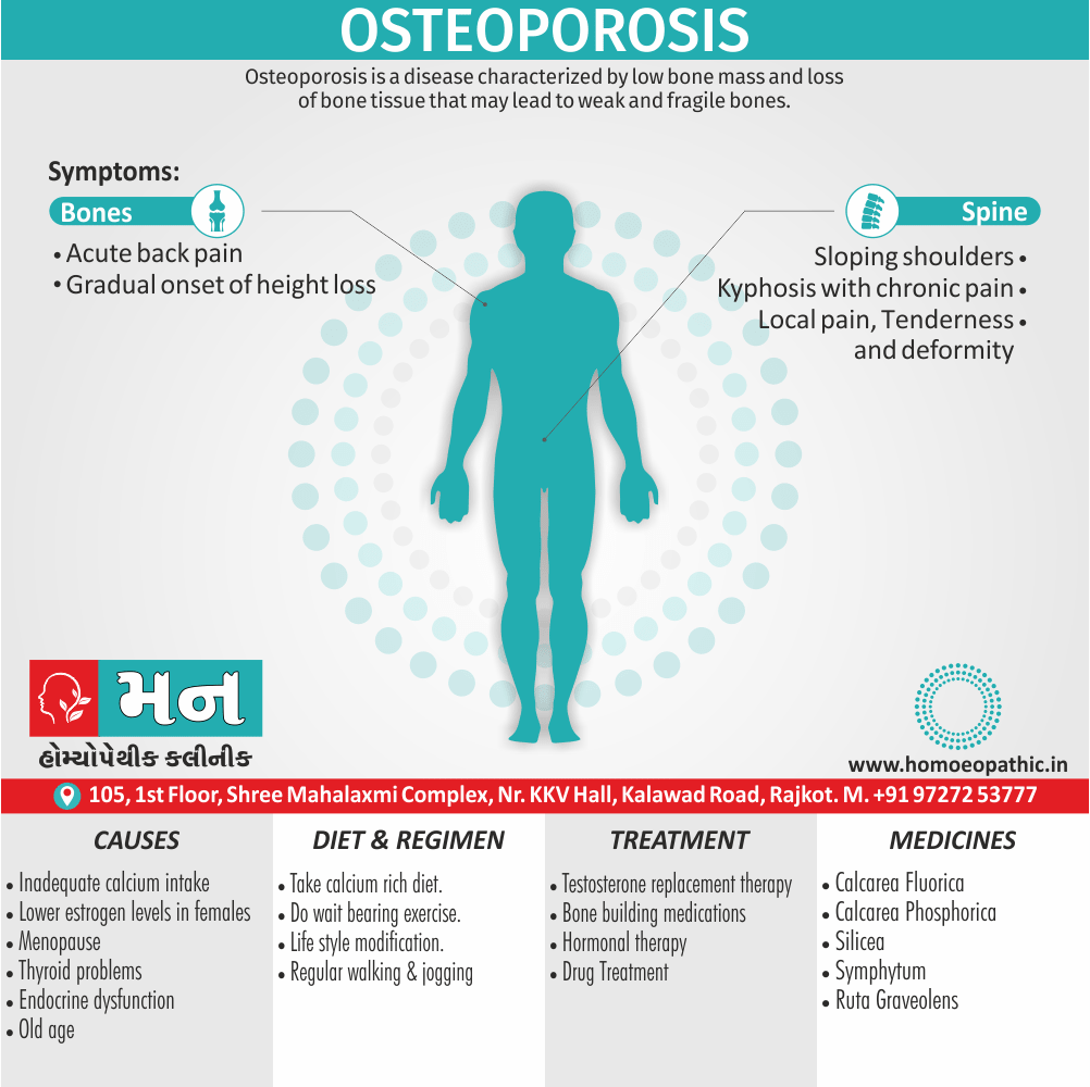
- Osteoporosis is an age-related disorder characterized by decreased bone mass and increased susceptibility to fractures of the hip, vertebra, and wrist. Furthermore, It occurs when bone resorption outpaces bone formation, since bone constantly undergoes cycles of resorption and formation (in other words, remodeling) to maintain the concentration of calcium and phosphate in the extracellular fluid. Signs of osteoporosis are vertebral compression, loss of body height, development of kyphosis, and hip fracture.
- Osteopetrosis is an abnormally dense bone, obliterating the marrow cavity, due to defective resorption of immature bone.
II. Joints in Skeletal System:
places of union between two or more bones.
Innervated as follows: The nerve supplying a joint also supplies the muscles that move the joint and the skin covering the insertion of such muscles (Hilton’s law).
Classified on the basis of their structural features into fibrous, cartilaginous, also synovial types.
A. Fibrous Joints (Synarthroses)
■ Joined by fibrous tissue, have no joint cavities, also permit little movement.
1. Sutures i.e.
■ In brief, Connected by fibrous connective tissue and found between the flat bones of the skull.
2. Syndesmoses i.e.
■ Connected by fibrous connective tissue.
■ Occur as the inferior tibiofibular and tympanostapedial syndesmoses.
B. Cartilaginous Joints
■ United by cartilage and have no joint cavity.
1. Primary Cartilaginous Joints (Synchondroses) i.e.
■ United by hyaline cartilage and permit little to no movement but allow for growth in length during childhood and adolescence.
■ Include epiphyseal cartilage plates (the union between the epiphysis and the diaphysis of a growing bone) and sphenooccipital and manubriosternal synchondroses.
2. Secondary Cartilaginous Joints (Symphyses) i.e.
■ Joined by fibrocartilage and are slightly movable joints.
■ Include the pubic symphysis and the intervertebral disks.
C. Synovial (Diarthrodial) Joints
■ Permit a great degree of free movement and classified according to the shape of the articulation and/or the type of movement.
■ Characterized by four features: joint cavity, articular (hyaline) cartilage, synovial membrane (which produces synovial fluid), also articular capsule.
1. Plane (Gliding) Joints i.e.
■ United by two flat articular surfaces and allow a simple gliding or sliding of one bone over the other.
■ Occur in the proximal tibiofibular, intertarsal, intercarpal, intermetacarpal, carpometacarpal, sternoclavicular, also acromioclavicular joints.
2. Hinge (Ginglymus) Joints i.e.
■ Resemble door hinges also allow only flexion and extension.
■ Occur in the elbow, ankle, also interphalangeal joints.
3. Pivot (Trochoid) Joints i.e.
■ Formed by a central bony pivot turning within a bony ring and allow only rotation (movement around a single longitudinal axis).
■ Occur in the superior and inferior radioulnar joints and in the atlantoaxial joint.
4. Condylar (Ellipsoidal) Joints i.e.
■ Have two convex condyles articulating with two concave condyles. (The shape of the articulation is ellipsoidal.)
■ Allow flexion and extension and occur in the wrist (radiocarpal), metacarpophalangeal, knee (tibiofemoral), and atlantooccipital joints.
5. Saddle (Sellar) Joints i.e.
■ Resemble a saddle on a horse’s back and allow flexion and extension, abduction and adduction, and circumduction but no axial rotation.
■ Moreover, Occur in the carpometacarpal joint of the thumb and between the femur and patella.
6. Ball-and-Socket (Spheroidal or Cotyloid) Joints i.e.
■ Formed by the reception of a globular (in other words, ball-like) head into a cup-shaped cavity and allow movement in many directions.
■ Besides this, Allow flexion and extension, abduction and adduction, medial and lateral rotations, and circumduction, and occur in the shoulder and hip joints.
CLINICAL ANATOMY
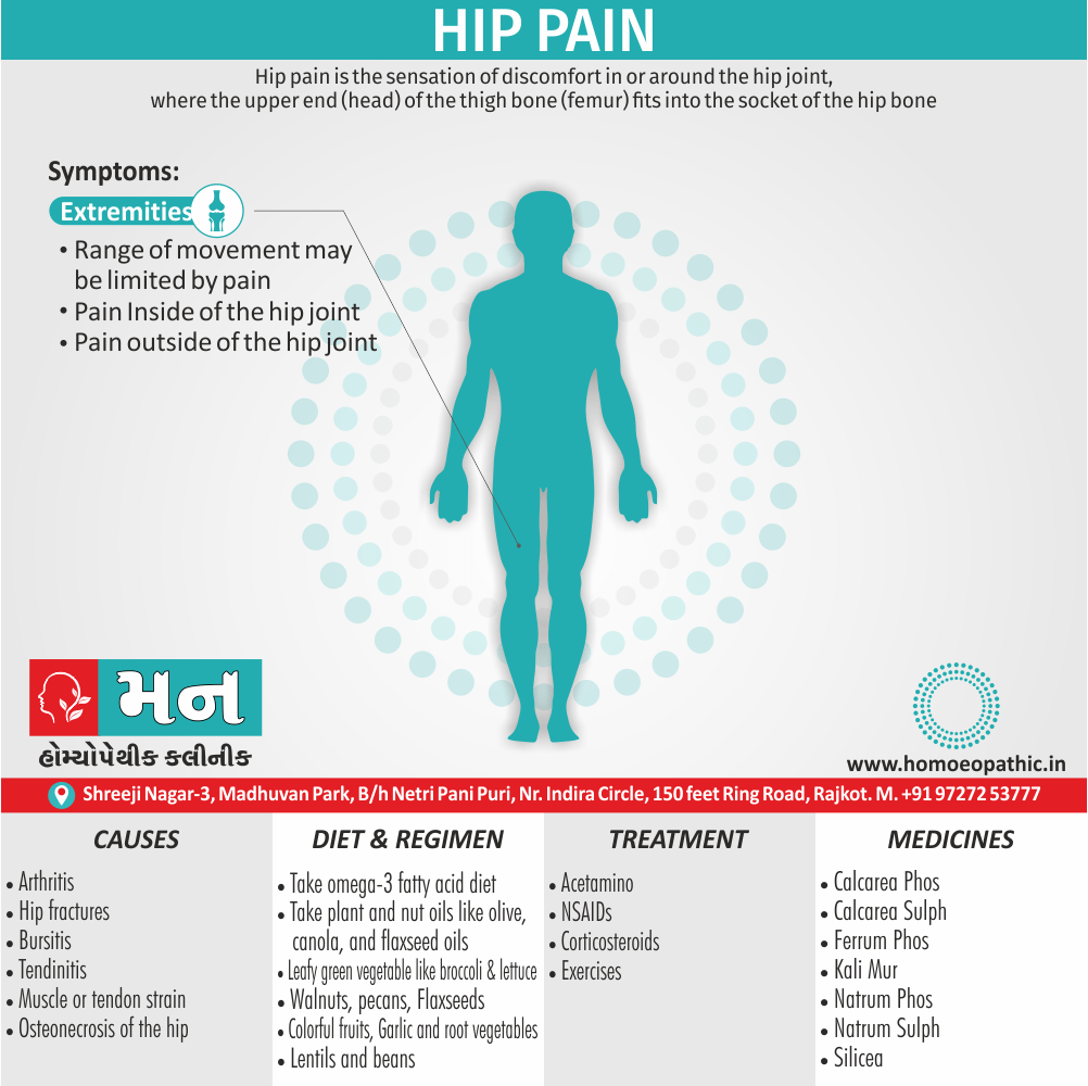
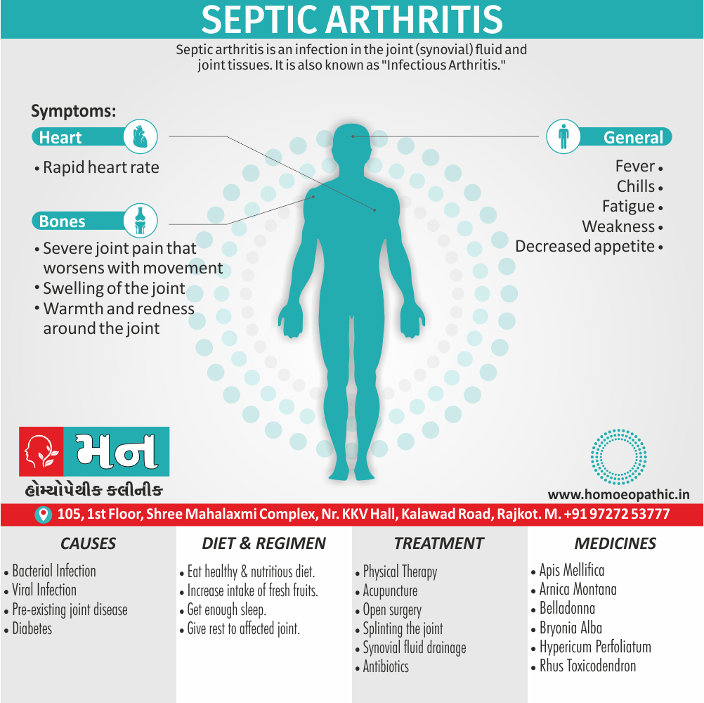
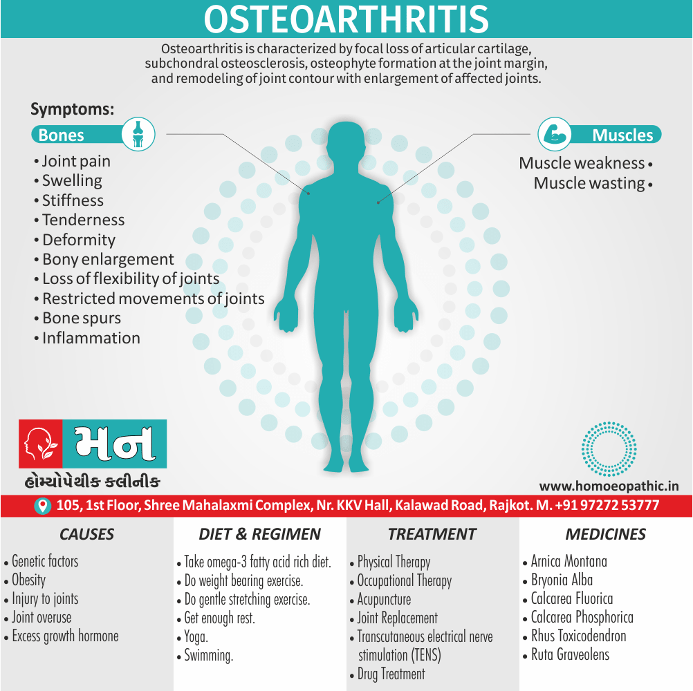
- Osteoarthritis is a noninflammatory degenerative joint disease characterized by degeneration of the articular cartilage and osseous outgrowth at the margins. It results from wear and tear of the joints; additionally commonly affects the hands, fingers, hips, knees, feet, and spine; and is accompanied by pain and stiffness.
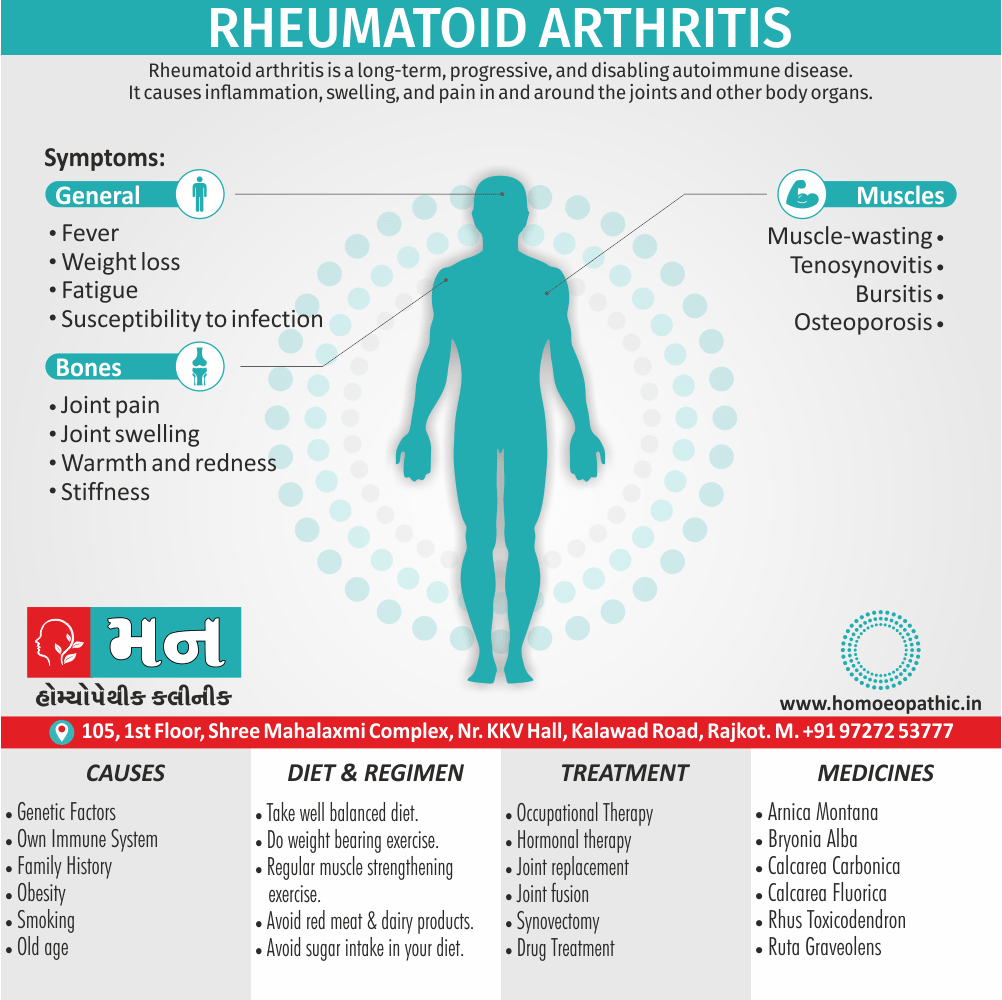
- Rheumatoid arthritis is an inflammatory disease primarily of the joints. It is an autoimmune disease in which the immune system attacks the synovial membranes and articular structures, leading to deformities and disability. Rheumatoid arthritis is most common symptoms are joint swelling, stiffness, also pain.
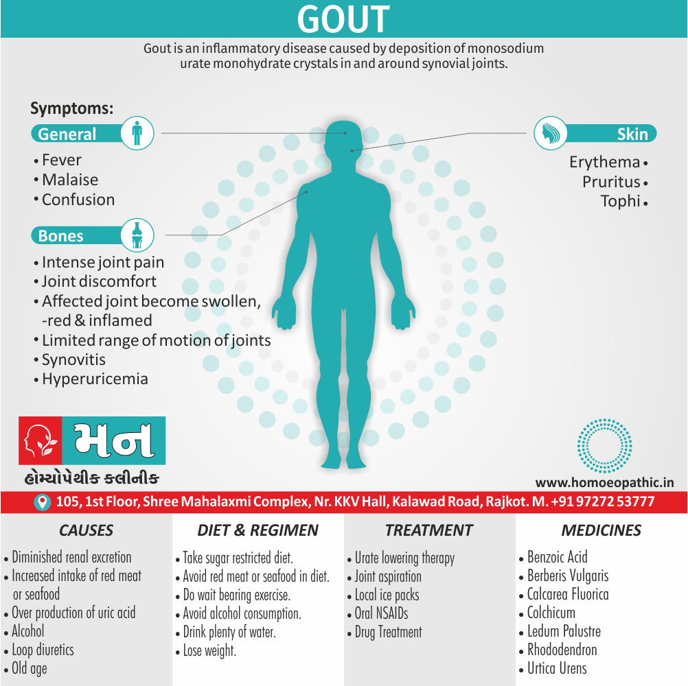
- Gout is a painful form of arthritis and caused by too much uric acid in the blood. Uric acid crystals are deposited in and around the joints, causing inflammation and pain, heat, redness, stiffness, tenderness, and swelling of the joint tissues.