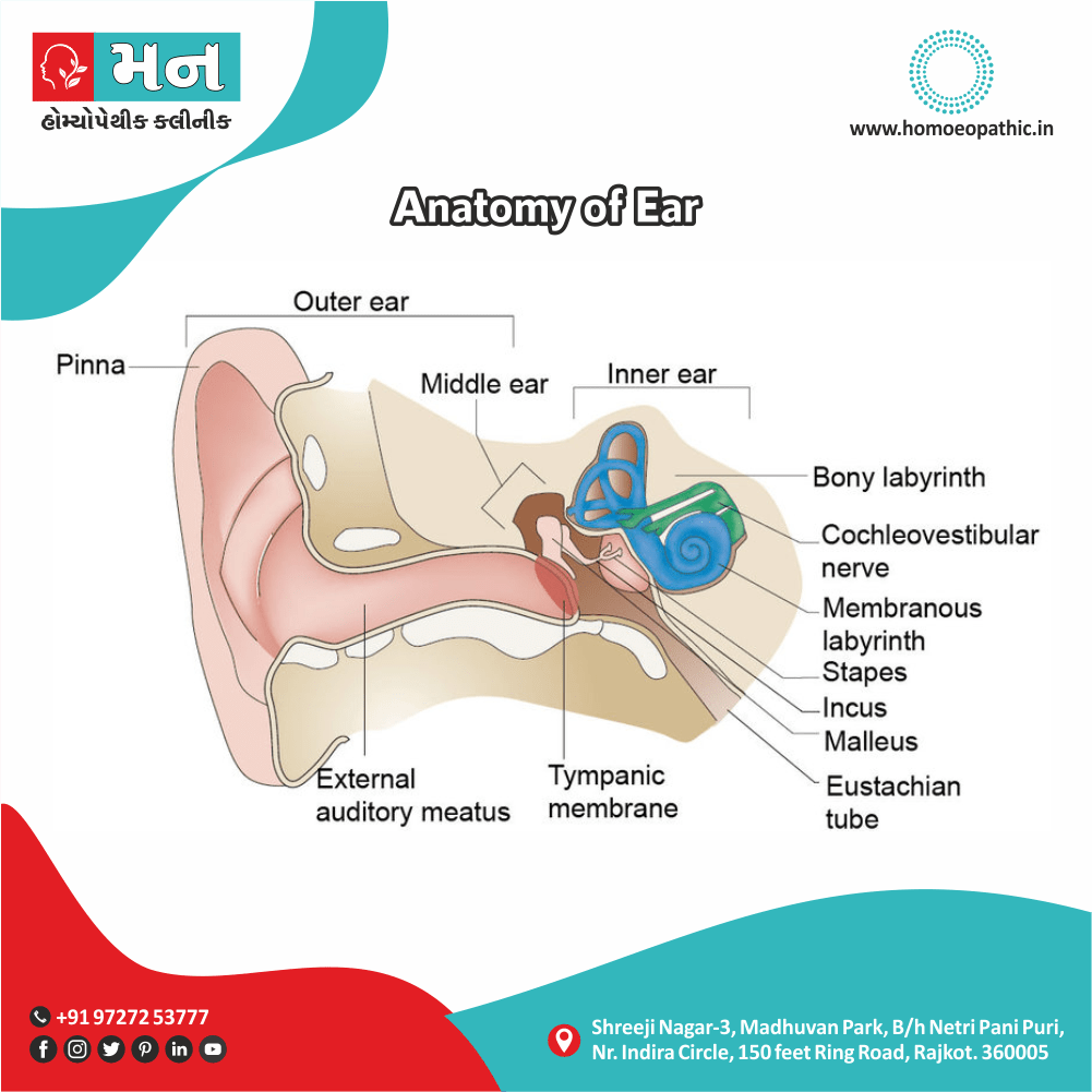
Anatomy of Ear:
The ear is divided into three part i.e.:
- Firstly, External ear
- Secondly, Middle ear
- Lastly, Internal ear or the labyrinth
1. The Anatomy of External Ear:
In Anatomy of External Ear consist of mainly 3 structures i.e.:
- Auricle or pinna,
- External acoustic canal
- Tympanic membrane
Auricle or Pinna in External Ear:
It is the first part of Anatomy of External ear. Basically, the entire pinna except its lobule and the outer part of external acoustic canal made up of a framework of a single piece of yellow elastic cartilage covered with skin. On the other hand, the latter is closely adherent to the perichondrium on its lateral surface while it is slightly loose on the medial cranial surface. Additionally, The various elevations and depressions seen on the lateral surface of pinna.
There is no cartilage between the tragus also crus of the helix, and this area is call incisura terminalis (Fig. Ear The auricular cartilage.). Moreover, An incision made in this area will not cut through the cartilage and is using for endaural approach in surgery of the external auditory canal or the mastoid. Pinna is also the source of several graft materials for the surgeon. Cartilage from the tragus, perichondrium either from the tragus or concha and fat from the lobule are frequently use for reconstructive surgery of the middle ear. Additionally, The conchal cartilage has also using to correct the depress nasal bridge while the composite grafts of the skin and cartilage from the pinna are sometimes used for repair of defects of nasal ala.
External Acoustic Canal in External Ear:
It extends from the bottom of the concha to the tympanic membrane also measures about 24 mm along its posterior wall. It is not a straight tube because its outer part is directing upwards, backwards and medially while its inner part is directing downwards, forwards and medially. Therefore, to see the tympanic membrane, the pinna has to pull upwards, backwards and laterally so as to bring the two parts in alignment.
The canal is dividing into two parts:
- Cartilaginous and
- Bony.
CARTILAGINOUS PART
It forms outer one-third (8 mm) of the canal. Cartilage is a continuation of the cartilage which forms the framework of the pinna. It has two deficiencies—the “fissures of Santorini” in this part of the cartilage and through them the parotid or superficial mastoid infections can appear either in the canal or vice versa. The skin covering the cartilaginous canal is thick and contains ceruminous and pilosebaceous glands which secrete wax. Hair is only confined to the outer canal and therefore furuncles (staphylococcal infection of hair follicles) are seen only in the outer onethird of the canal.
BONY PART
It forms inner two-thirds (16 mm). Skin lining the bony canal is thin and continuous over the tympanic membrane. It is devoid of hair also ceruminous glands. Especially, 6 mm lateral to tympanic membrane, the bony meatus presents a narrowing called isthmus. Generally, Foreign bodies, lodged medial to the isthmus, get impacted, and are difficult to remove. Anteroinferior part of the deep meatus, beyond the isthmus, presents a recess called anterior recess, which acts as a cesspool for discharge and debris in cases of external and middle ear infections (Figure 1.2). Anteroinferior part of the bony canal may present a deficiency (foramen of Huschke) in children up to the age of four or sometimes in adults, permitting infections to and from the parotid.
Tympanic Membrane in External Ear:
It forms the partition between the external acoustic canal also the middle ear.
Tympanic Membrane obliquely set and as a result, its posterosuperior part is more lateral than its anteroinferior part. It is 9–10 mm tall, 8–9 mm wide and 0.1 mm thick. Tympanic membrane can divide into two parts i.e.:
-
Firstly, PARS TENSA
-
Secondly, PARS FLACCIDA (SHRAPNELL’S MEMBRANE)
a. PARS TENSA
It forms most of tympanic membrane. Its periphery thicken to form a fibrocartilaginous ring called annulus tympanicus, which fits in the tympanic sulcus. Moreover, The central part of pars tensa is tenting inwards at the level of the tip of malleus and is called umbo. A bright cone of light seen radiating from the tip of malleus to the periphery in the anteroinferior quadrant
b. PARS FLACCIDA (SHRAPNELL’S MEMBRANE)
This situate above the lateral process of malleus between the notch of Rivinus and the anterior and posterior malleal folds (earlier called malleolar folds). It is not so taut and may appear slightly pinkish. Various landmarks seen on the lateral surface of tympanic membrane are shown in Figure 1.4.
LAYERS OF TYMPANIC MEMBRANE
Tympanic membrane consists of three layers i.e.:
Outer epithelial layer
which is continuous with the skin lining the meatus.
Inner mucosal layer,
which is continuous with the mucosa of the middle ear.
Middle fibrous layer
which encloses the handle of malleus and has three types of fibres—the radial, circular also
parabolic (Figure 1.5).
Fibrous layer in the pars flaccida is thin and not organized into various fibres as in pars tensa.
RELATIONS OF EXTERNAL ACOUSTIC MEATUS
- Superiorly: Middle cranial fossa Triangular fossa Helix Spine of helix Tail of helix Antitragus
- Posteriorly: Mastoid air cells and the facial nerve
- Inferiorly: Parotid gland
- Anteriorly: Temporomandibular joint
Posterosuperior part of deeper canal near the tympanic membrane is related to the mastoid antrum. “Sagging” of this area may be noticed in acute mastoiditis.
PINNA
1. Greater auricular nearve (C2,3) supplies most of the
medial surface of pinna and only posterior part of the
lateral surface (Figure 1.6).
2. Lesser occipital (C2)supplies upper part of medial surface.